


Combination of Liver and brain hydatidosis: A rare presentation
Contributed by
Dr Syed Aalishan Fatima & Prof. Majid Jahangir
Dept. of Radiodiagnosis
Govt Medical College Srinagar ,Kashmir
Clinical scenario:
A 60 year old female patients presented with history of pain abdomen ,breathlessness ,
headache .She also complained of vomiting for 2day duration
With these symptoms she was admitted to emergency ward of our hospital.
O/E she was conscious , oriented and her vitals were stable
The neck was free and there were no meningeal signs
Initial NCCT showed a calcified mass in the left frontoparietal with profound oedema .

Later MRI brain was done which showed the lesion was isointense to cerebral white matter on T1-weighted images.

T2- weighed images revealed a heterogeneous low-intensity mass with peripheral high signal intensity consistent with oedema , blooming and peripheral ring enhancement on post contrast study
Contrast enhanced CT scan abdomen showed a non enhancing infiltrating hepatic solid mass that contains multiple cystic-necrotic hypodense areas and hyperattenuating foci of internal calcifications .
Infiltration into hepatic veins and intrahepatic IVC was also noted.
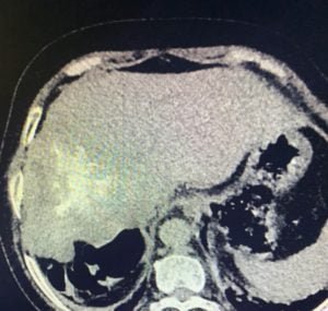
Fig CT scan abdomen showing liver mass
Teaching message :
It is known that human parasitic zoonoses sometimes cause diagnostic and therapeutic problems. Radiologic imaging findings may contribute to the diagnosis of alveolar hydatid disease. AE lesions may be misdiagnosed as malignancies, and it is important for radiologists to have good command of typical imaging features of AE and to provide early diagnosis for patients from high prevalence areas, thereby guiding clinicians in the correct direction.
Further reading
Hydatid disease of the brain and spine
Join the mailing list!
Get the latest articles delivered right to your inbox!