

Think out of the box , Man with low backache
A 58 year old male presented with history of low back ache of 6 weeks duration without any history of trauma .He was able to do his activities of daily life ,nevertheless with difficulties due to disabling pain.
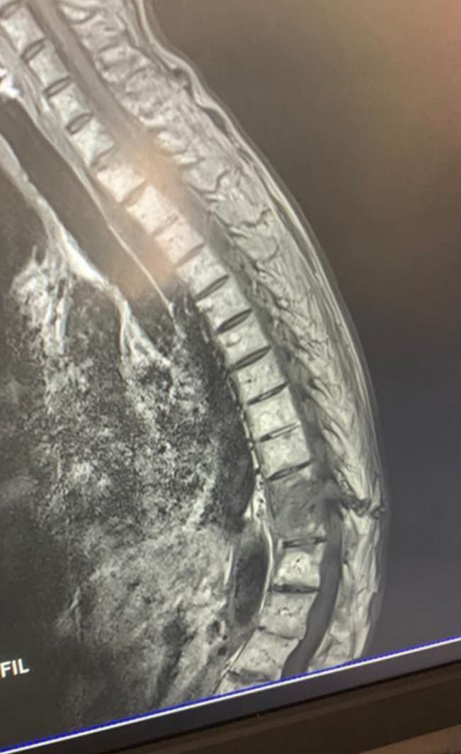
Apart from baseline investigations he was advised MRI spine as shown in Fig1
MRI showed involvement of 2 adjacent vertebral body and disc with prevertebral soft tissue mimicking spinal TB
The patient was labelled as spinal TB on the basis of MRI .
ATT was started but he had no relief .

However, a careful observation of MRI shows facet joint fusion at several levels in addition to fracture through vertebro discal junction is noted at the same level as well.
Raising the possibility of Ankylosing spondylitis with Anderson lesion.
Andersson lesion is a rare aseptic discovertebral lesion occurring in individuals with ankylosing spondylitis. It should be differentiated from infective spondylodiscitis and metastatic disease by its classical appearance on radiographs and MRI.It often requires surgical management to alleviate pain, and prevent it leading to pseudoarthrosis or kyphosis.

As such Plain X Ray and CT was advised which showed classic bamboo spine and Anderson lesion as shown in
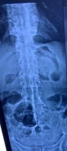
The final diagnosis was Ankylosing spondylitis .
Further reading : Click the Link. Ankylosing Spondylitis Imaging
View in browser :The Health Guide
Join the mailing List of The Health Guide
Join the mailing list!
Get the latest articles delivered right to your inbox!