


Case of the month :Mongolian spots in a newborn
Contributed by
Dr.Ghulam Nabi, MD.
Pediatric Consultant and Neonatologist.
Bugshan Hospital Jeddah, Saudi Arabia
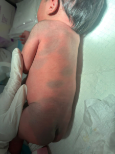
Clinical scenario: Full term female baby born to 27 years old Yemeni mother, gravida two Para one was presented to our clinic with spots on the back. Clinical history and examination: Born after a normal vaginal delivery. The baby had a Apgar score 7 and 9 at 1 and 5 minutes, Examination The baby was alert weight 2.7 kilogram. Head circumference 33 centimeters, length 49 cm. Neonatal reflexes present. Feeding normal .The baby had passed first stool (meconium). Clinically all systems were normal The skin examination showed extensive light blackish discoloration of the skin at shoulders. back, buttocks, both upper limbs and both lower limbs more on left lower limb (Figure 1). These are called Mongolian spot (Dermal Melanocytosis).
Congenital dermal Melanocytosis (previously called Mongolian spots) is the most common birth mark. These are slate grey, dark blue or purple bruise like macular spots and have variable defined margins, usually located over the sacrum. These macules or patches are occasionally noted over the shoulders and back. The spots may be solitary or numerous and often involves larger areas. Documentation is important because it can be confused with bruising after discharge. The incidence of these lesions, most common in African American, Asian and Hispanic infants’ 25-80%. The peculiar hue of these macules is a result of dermal location of melanin containing melanocytes that are presumably arrested in their migration from neural crest to epidermis. They usually fade during the first few years of life as a result of darkening of overlying skin. If lesions persist, they may be treated with lasers if desired. Malignant degeneration does not occur. Rarely Mongolian Spots are associated with Hurler or Hunter syndromes, Gangliosidosis, Nieman Pick disease, Mucolipidosis and Manosidosis. Dermal Melanocytosis in the area of first and second division of the trigeminal nerve is called Nevus of Ota. References
|
Join the mailing list!
Get the latest articles delivered right to your inbox!