Sister Mary Joseph nodule revisited !
Clinical scenario:
A 65 yr old female presented with a discharging swelling in the umbilical area of 2 months duration .She had been to a local doctor who prescribed some antibiotics but the lesion continued to grow and didn’t heal .She had no jaundice , itching but complained of anorexia and weight loss of 3 Kg in the same period.
Examination:
She was conscious , oriented hemodynamically stable and had no icterus or lymphadenopathy. Her abdominal examination revealed an umbIlical nodule around 4cms with serosanguinous discharge and it had firm margins Fig 1
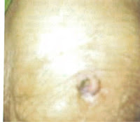 |
| Fig 1 UmbIlical nodule SJN |
Her abdominal examination revealed hepatomegaly 2 cms below costal margin .There was no ascites .Her systemic examination was normal .
Evaluation:
The hemogram revealed hemoglobin of 10.8 gm/dl and her TLC and platelets were normal . Her renal functions and liver functions were normal .Keeping in view non healing nature of this umbilical swelling , biopsy was done from the edge of the nodule which showed metastatic adenocarcinoma .Fig2 .Hence the nodule proved to be sister Mary Joseph nodule .
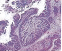 |
| Fig 2 Biopsy from umbilical nodule |
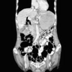
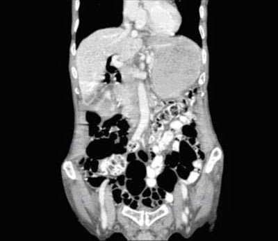
Later CT scan abdomen was done Fig 3 which revealed gallbladder mass .Patient had an inoperable carcinoma of gall bladder and she later developed progressive cholestasis and deep Jaundice and succumbed to her illness unfortunately .
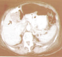 |
| Fig 3 CT abdomen showing carcinoma Gallbladder (arrow ) |
Teaching message :
“Non healing ulcers must be biopsied” .
Umbilical metastasis is named Sister Mary Joseph nodule (SJN) The name is after the vigilant head nurse working in Mayo clinic nearly a century ago .She observed patients with this findings were having advanced malignancy . Her observation was published in 1949 in Hamilton Baily .
Men with SJN usually have GI malignancies while women with SJN have ovarian malignancies . It can occasionally occur due to lung malignancies even.
The size may range few cms and even as long as 10 cms have been recorded . This finding reflects metastasis through lymphatic channels or through peritoneum or through embryological remnants .
Further Reading: Gall bladder cancer
Join the mailing list!
Get the latest articles delivered right to your inbox!
