


Case of the month :A hidden spurter: proximal gastric Dieulafoy lesion missed on two endoscopies, rebleeding after clipping and treated by endoscopically guided wedge resection
Contibuted by :
Dr Mahmoud Elkaramany,
Consultant Gastroenterologist Cardiff and Vale Univeristy Health Board UK
A 64-year-old woman (Mrs X) presented with recurrent episodes of melaena.
On each admission her Hb fell to about 60 g/L, requiring blood transfusion.,She denied any use of NSAIDs and was not on blood thinners
OGDs on the first two admissions showed no bleeding source.
On the third admission she presented with history of melaena of 3 days duration .
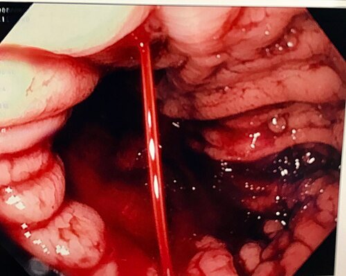
At the third OGD, performed by the author, a small lesion on the lesser curve of the proximal stomach began to spurt arterial blood vigorously as the scope approached (Forrest Ia). There was no surrounding ulceration, consistent with a Dieulafoy lesion.
Two endoclips were placed across the vessel, followed by injection of diluted adrenaline (1:10,000) around the site. Complete haemostasis was achieved.. She completed three days of intravenous proton pump inhibitor (PPI) and was discharged well, with no further melaena. CT angio did not show any bleeding .Note was made of adrenal incidanteloma and urology advice was followed
However, within days of discharge she re-presented with melaena, leading to repeat OGD and then surgery.
Table 1. Clinical timeline
| Stage | Event | Key findings / action |
| 1 | First admission | Melaena, Hb ~60 g/L; OGD 1 non-diagnostic |
| 2 | Second admission | Melaena, Hb ~60 g/L; OGD 2 non-diagnostic |
| 3 | Third admission | OGD 3: spurting Dieulafoy lesion, proximal lesser curve; 2 clips + adrenaline; CTA no active bleeding |
| 4 | Discharge | Well after 3 days of IV PPI |
| 5 | Readmission | Melaena; OGD 4: old blood, adherent clot over clipped site |
| 6 | Surgery | Endoscopically guided gastric wedge resection |
| 7 | Follow-up | Well, normal Hb, no further bleeding |
Exact dates are omitted to protect patient anonymity.
Endoscopic findings and haemostasis
At readmission, OGD showed a large amount of old blood and clot in the stomach. Both clips remained in place, with a large adherent clot over the treated site. She was managed with an intravenous PPI infusion, a prokinetic to clear the stomach, serial Hb monitoring and repeat imaging. Given rebleeding despite combined endoscopic therapy, definitive surgical resection was planned.
Surgery and outcome
She underwent endoscopically guided gastric wedge resection. The short gastric vessels were divided and the stomach mobilised to expose the proximal stomach and lesser curve.
Intraoperative OGD located the lesion using the previously placed clips. Sleeve gastrectomy and fundectomy were considered, but wedge resection of the clip-marked area was chosen as the least extensive option. An anterior gastrotomy was made and the clipped area delivered through it. The wedge was resected with an Echelon linear stapler (green cartridges, three firings). Stay sutures were released and the gastrotomy closed with two further firings.
Completion endoscopy showed the expected mild narrowing of the mid-body, with a largely patent lumen and satisfactory haemostasis.
She recovered and was discharged home. At follow-up she was well, with no further melaena and a normal Hb. The adrenal incidentaloma remains under urology follow-up.
Discussion
This case shows how a Dieulafoy lesion can hide in plain sight. Two OGDs failed to find the source despite repeated severe bleeds; the lesion was seen only when it was actively bleeding.
Why it is missed. The lesion is tiny, lacks surrounding ulceration and bleeds intermittently. In the proximal stomach it may sit in a blind spot or be covered by clot or pooled blood. Careful retroflexed views of the fundus and cardia, water irrigation, a distal cap and patient repositioning all improve detection [3,4]. Early endoscopy, and repeat endoscopy during a fresh bleed, raise the yield. Where endoscopy is repeatedly negative, CT angiography or mesenteric angiography can localise an actively bleeding vessel [4].
Endoscopic treatment. Endoscopic therapy achieves primary haemostasis in over 90% of cases [2,5]. Adrenaline injection alone carries a high rebleeding risk, so combination therapy is preferred. Mechanical methods such as clips or band ligation appear more durable than thermal or injection therapy alone [5,6]. Our combination of clips to occlude the vessel and adrenaline injection is in line with this.
Role of surgery. Rebleeding after endoscopic treatment occurs in about 10–20% [5]. Repeat endoscopic therapy or angiographic embolisation are usual next steps, with surgery reserved for failure or selected high-risk cases [2]. In our patient, the large adherent clot over the clipped site and her history of repeated bleeds with Hb near 60 g/L favoured definitive resection.
The main surgical difficulty is finding a small lesion from the serosal side. Here the endoscopic clips served as markers, and intraoperative endoscopy through a combined approach allowed a limited wedge resection instead of fundectomy or sleeve gastrectomy. The resulting mid-body narrowing was expected and did not compromise the lumen.
Learning points
- Consider a Dieulafoy lesion in any patient with recurrent, severe UGIB and a non-diagnostic OGD.
- Most lie in the proximal stomach; a careful retroflexed look at the fundus and cardia is essential.
- Endoscopy during or soon after an active bleed gives the best chance of finding the lesion.
- Combination therapy (mechanical clips plus adrenaline) is preferred over adrenaline alone.
- Surgical wedge resection offers definitive treatment when bleeding recurs or endoscopic control is uncertain.
References
- Lee YT, Walmsley RS, Leong RW, Sung JJ. Dieulafoy’s lesion. Gastrointest Endosc. 2003;58(2):236–243.
- Baxter M, Aly EH. Dieulafoy’s lesion: current trends in diagnosis and management. Ann R Coll Surg Engl. 2010;92(7):548–554.
- Nojkov B, Cappell MS. Gastrointestinal bleeding from Dieulafoy’s lesion: clinical presentation, endoscopic findings, and endoscopic therapy. World J Gastrointest Endosc. 2015;7(4):295–307.
- Jeon HK, Kim GH. Endoscopic management of Dieulafoy’s lesion. Clin Endosc. 2015;48(2):112–120.
- Chung IK, Kim EJ, Lee MS, et al. Bleeding Dieulafoy’s lesions and the choice of endoscopic method: comparing the hemostatic efficacy of mechanical and injection methods. Gastrointest Endosc. 2000;52(6):721–724.
- Park CH, Sohn YH, Lee WS, et al. The usefulness of endoscopic hemoclipping for bleeding Dieulafoy lesions. Endoscopy. 2003;35(5):388–392.
Join the mailing list!
Get the latest articles delivered right to your inbox!


