


2 mins read
Vacuum extraction as a treatment modality of neonatal skull depression !
Clinical scenario:
Twin infants were born at 39 weeks of gestation to a healthy multiparous lady in our hospital. She had no history of any known trauma during the pregnancy or at delivery. Her gestational course was uncomplicated .
The antenatal ultrasound in the third trimester revealed twin pregnancy and normal maternal uterus,her delivery of twin B was by assisted breech.
Twin A was a male baby, birth weight 1.9 kg.and was discharged home on the second day of admission.Twin B was admitted for management of skull depression .
Examination of the Twin B:
Twin B was also a baby boy.
APGARSCORE ( Appearance, Pulse, Grimace, Activity,and Respiration.) was 7 and 9 at 1 and 5 minutes. The birth weight was 2.7 kg. His head circumference was 33.5 cm, and length 50 cm.
The vital signs were stable, He had no dysmorphic features and he was active .He had normal primitive neonatal reflexes (Watch video demonstration of primitive reflexes ) and his neurological examination was also normal.
However, head examination showed that the anterior fontanel was flat, not bulging, and the left parietal bone was depressed approximately 4 cm length and 3 cm width (Figure 1).
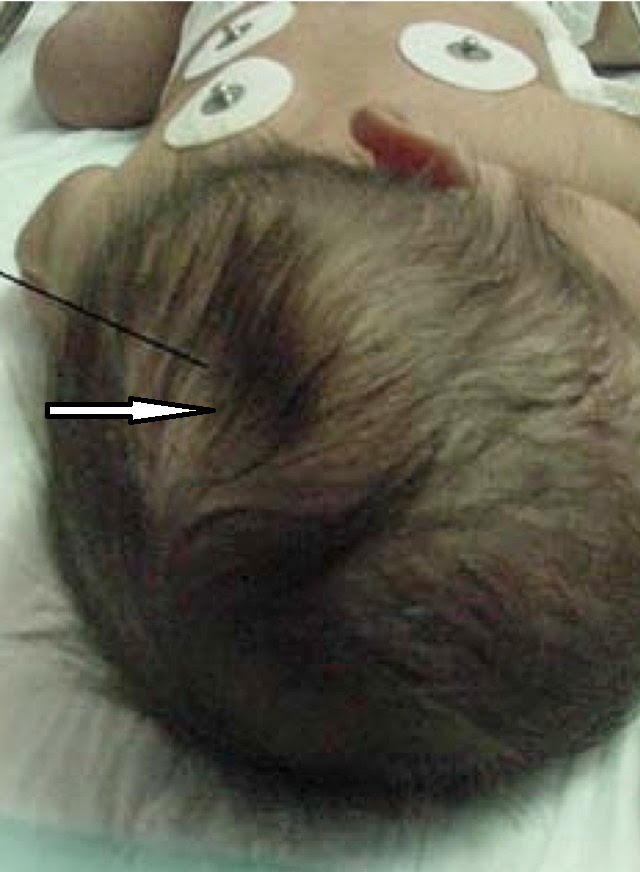 |
| Fig 1 Arrows showing depression in skull |
There was no edema or hematoma in this area. He was transferred to the neonatal ICU for further evaluation.
Evaluation and management:
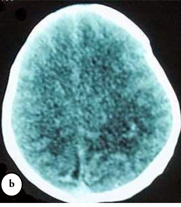
Complete blood count, prothrombin time, and partial prothrombin time were normal for age. The CT brain was obtained and showed skull depression of the left parietal area (Figure 2).
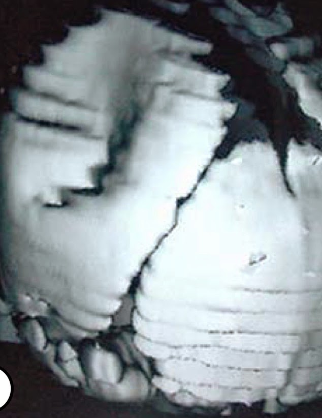 |
| Fig 2 CT scan showing three dimensional view of depression |
He remained asymptomatic, and the depressed fracture was successfully elevated by obstetrical vacuum extractor. (Read more about Vaccum extraction procedure .)
On the second day after birth, he was in good condition, good oxygen saturation in room air, feeding was started and well tolerated. The vital signs were stable. On day 3, repeat CT scan showed corrected depressed fracture
(Figures 3)
 |
| Fig 3 Repeat CT scan head post skull elevation by Vacuum. |
On the sixth day after birth, he was discharged home in good condition .
Teaching message :
Congenital depression of the fetal skull are rare lesion occurring in 1-2.5 per 10,000 births resulting in asymmetrical skull. This depression is caused by exaggerated or prolonged pressure applied to the fetal head in utero or during delivery. The management regimen for depressed skull fractures in infants can be conservative or surgical. Conservative management is a reasonable treatment option in uncomplicated cases .
Further reading :
Vaccum extraction as a treatment modality of neonatal skull depression
Contributed by:
Dr. Adnan Amin Alsulaimani.
Professor of Neonatology.
Dean of College of Medicine,
Taif University .KSA.
Join the mailing list!
Get the latest articles delivered right to your inbox!