Kidney cancer: Causes and a new tool in it’s diagnosis.
Contributed by
Abeer Al-Hussaini, Ghulam Nabi*
Address for correspondence
Professor Ghulam Nabi
Professor of Surgical Uro-oncology and Consultant Robotics Surgeon
Ninewells Hospital, Dundee, UK
Kidney cancer develops from cells lining the tubules of the organ and is the 9th most common cancer type in females and the 7th most common type of cancer in males.
Most commonly it affects only one kidney , but on occasions both kidneys may harbor this at the same time (synchronous) or in succession (metachronous).
The disease, in particular in its early stages, does not have any symptoms.
When someone does have symptoms, and usually diagnosed by scans (ultrasound, CT, MRI) of the abdomen for unrelated reasons.
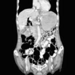
By and large, it appears as solid mass on imaging but sometimes it can be seen as cystic (1). Later has a good prognosis.
The most common symptoms observed in kidney cancer are blood in the urine (2)and or a lump in the flank area.
There are a number of risk factors which potentially can increase your chance of getting a disease. Having a risk factor/s does not mean having a disease but it is emphasized that one has to be aware of. The risk is increased due to:
- Smoking
- Increase in weight (obese)
- High blood pressure
- Gender – higher risk in men as compared to women.
- Chronic kidney disease and being on dialysis treatment.
- History of family members with kidney cancer
- Chronic use of medication: pain-relieving drug called phenacetin.
- Certain abnormal disorders due to faulty genes, such as von Hippel-Lindau disease, Birt Hogg Dube syndrome, and others
- Chronic exposure to asbestos or cadmium
- Surgery remains the mainstay of treatment for localized and locally advanced disease and in those with cancer spread to other parts of the body, tyrosine kinase inhibitors and check-point inhibitors are offered. In general, this tumor is resistant to chemo and radiotherapy. In selected cases, surgery can be offered in the context of overall planning in patients with metastatic disease (cytoreductive nephrectomy,
New tools in it s diagnosis :
Kidney cancer incidence is increasing partly due to increased use of ultrasound and CT scan for various conditions. However, when the cancer in kidney is small (less than 4cm), there may be chance that this may be benign. Again, this may not be known to physicians unless they interpret this through biopsies. The procedure of biopsies is painful one and may not provide level of finality most would be expecting from this.
Abeer and colleagues through an exhaustive research provided a solution to interpretation of CT scan of patients affected by kidney cancer.
They used radiomics approach to glean out data from images of kidney cancer which is not visible to human eyes .
The data included minute details such as make-up of cancer tissue, just like interpretation of bricks (pixel in images) in the walls of a house.
The data was then subjected to various mathematical algorithms to make distinctions between various regions of cancers and with attempts to know which areas are more aggressive than others.
The paper has shown that using this approach, a more precise diagnosis of kidney cancer and its grade (marker of its aggressivity and marker of its killing power) in contrast to traditional approach.
A significant advantage of this approach for clinicians is that it is quicker, non-invasive and results interpretation is not dependent on human eyes but through a quantitative approach using computers (machines).
The findings of research are the focus of further research and may involve multiple centers across the globe. A significant advancement achieved through this paper almost certainly pushes the boundaries of human eye aid diagnosis of not only kidney cancer but also a number of other conditions.
References
- Abdullah BJ, Ng KH (2001) In the eyes of the beholder: what we see is not what we get. Br J Radiol 74(884):675–676
- Milstein A, Adler NE (2003) Out of sight, out of mind: why doesn’t widespread clinical quality failure command our attention? Health Aff (Millwood) 22(2):119–127
- Graber M, Gordon R, Franklin N (2002) Reducing diagnostic errors in medicine: what’s the goal? Acad Med 77(10):981–992
- Robinson PJ (1997) Radiology’s Achilles’ heel: error and variation in the interpretation of the Röntgen image. Br J Radiol 70(839):1085–1098
Join the mailing list!
Get the latest articles delivered right to your inbox!

